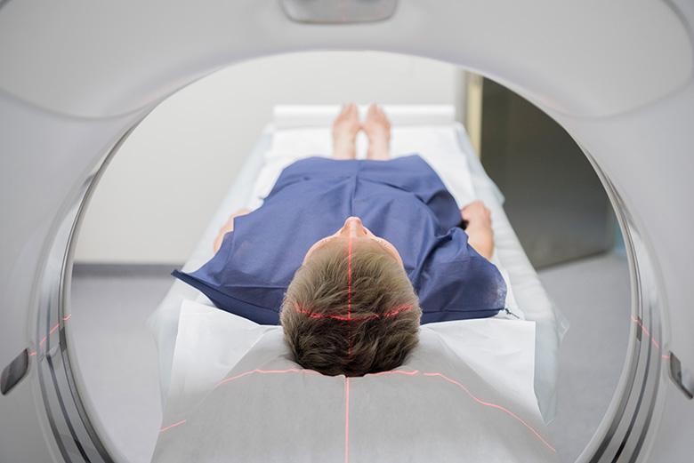
 man-taking-scan
A Positron Emission Tomography (PET) scan is an advanced medical imaging technique that offers detailed insights into the functional aspects of organs and systems within your body. This sophisticated scan is frequently combined with anatomical imaging techniques, such as a Computerized Tomography (CT) scan or a Magnetic Resonance Imaging (MRI) scan, performed simultaneously. By integrating these techniques, medical professionals gain the ability to:
- Assess the functional activity level of an organ or tissue based on its appearance in the scan.
- Detect abnormalities at a cellular level, often before changes in the organ’s shape become apparent.
PET scans are commonly utilized in the evaluation and diagnosis of cancer, neurological disorders affecting the brain, and cardiovascular conditions impacting the heart. When compared to MRI or CT scans alone, PET-CT or PET-MRI scans can provide a more comprehensive understanding, particularly for determining the stage of cancer. These scans are also valuable in assessing the effectiveness of cancer treatments. In cardiology, specialized Rubidium PET-CT scans can determine if the heart muscle is receiving adequate blood flow under various conditions.
How to Interpret PET Scan Results
During a PET scan, a radioactive tracer, known as a radiopharmaceutical, is introduced into the bloodstream. This tracer tends to accumulate in areas with higher levels of metabolic activity, such as diseased tissues. The PET scan then detects the emissions from this tracer to create multidimensional images of the body. Often, PET scans are integrated with CT scans, merging the structural details from the CT images with the functional information from the PET images.
The results of a PET scan are typically presented as a color-coded map. Areas with higher radiotracer concentration, indicating increased metabolic activity, will appear in brighter colors, often reds and yellows. Areas with lower activity will appear in darker or cooler colors, like blues and greens. Understanding these color variations is key to reading PET scan results.
Deciphering the PET Scan Image
While you will always need a specialist, like your doctor, to give you a definitive interpretation, understanding the basics of what you are seeing can be helpful. Here’s a simplified guide to reading PET scan results:
-
Areas of Increased Activity (Hot Spots): Bright colors (reds, oranges, yellows) on a PET scan typically indicate areas of increased metabolic activity. This could be due to various reasons, including:
- Tumors: Cancer cells often have a higher metabolism than normal cells, leading to increased tracer uptake and appearing as “hot spots.” PET scans are highly effective in detecting and staging many types of cancer.
- Inflammation: Areas of inflammation also exhibit increased metabolic activity and can show up as hot spots on a PET scan.
- Infection: Similar to inflammation, infections can cause increased metabolic activity in the affected area.
-
Areas of Decreased Activity (Cold Spots): Darker or cooler colors (blues, greens) usually represent areas with lower metabolic activity. This can indicate:
- Tissue Damage or Scarring: Areas of tissue damage or scarring might show reduced metabolic activity.
- Reduced Blood Flow: In cardiac PET scans, “cold spots” might indicate areas of reduced blood flow to the heart muscle.
-
Normal Activity: Areas showing intermediate colors, not excessively bright or dark, generally represent normal metabolic activity for that particular organ or tissue.
It’s important to note that interpreting PET scan results is not simply about identifying colors. The context of the patient’s medical history, the location and intensity of the tracer uptake, and comparison with other imaging modalities like CT or MRI are crucial for accurate interpretation. Radiologists and nuclear medicine physicians are specialists trained to analyze these complex images and provide detailed reports to your doctor.
Why is a PET Scan Necessary?
PET scans are valuable diagnostic tools because they can:
- Measure critical bodily functions such as blood flow, oxygen utilization, and glucose metabolism.
- Identify organs and tissues that are malfunctioning or inflamed.
- Detect cancerous tumor cells, aiding in assessing cancer spread (metastasis).
- Evaluate the effectiveness of cancer treatments by monitoring changes in metabolic activity.
PET scans can often detect cancers earlier than other imaging techniques like CT or MRI scans. They also help doctors determine the likelihood of cancer spreading and guide treatment strategies.
When Might You Need a PET Scan?
A doctor might recommend a PET scan in various situations, including:
- To detect potential cancer or determine its stage.
- To assess if vital organs, such as the heart, are receiving adequate blood flow in critical areas.
- To investigate suspected infections or inflammation within the body, including organs and bones.
- To examine organ abnormalities.
- To monitor the response to cancer treatment.
Furthermore, PET scans can assist in surgical planning for epileptic seizures by pinpointing the brain region triggering epilepsy. This technique is also used in the evaluation of neurological conditions like Alzheimer’s disease and Parkinson’s disease, as the images can reveal areas of the brain that are not functioning normally, allowing for earlier and more effective management.
Risks and Complications Associated with PET Scans
PET scans are generally considered safe and painless procedures. However, if you experience claustrophobia or fear of enclosed spaces, you may feel anxious about being inside the scanning machine. It is essential to inform your doctor about such concerns so they can determine the best approach to manage your comfort and ensure a successful scan.